Good treatment starts with seeing clearly.
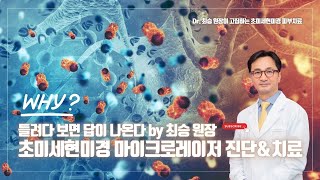
The Micro Laser System is Cheongdam Noel's concept for integrating close observation with laser treatment planning. It does not mean that one microscope determines a diagnosis. It means the clinic uses multiple visual inputs — including clinical examination, photography and high-magnification visualization — to understand the skin before selecting treatment.
Different lesions may share a similar color at first glance.
Looking more closely can help reveal differences in border, surface, thickness and visible vascular patterns — information that influences which laser to use and how.
High-magnification visualization within the laser workflow.
ZEISS TIVATO 700 is a surgical visualization system. At Cheongdam Noel, its high-magnification view is incorporated into selected laser workflows to observe the treatment field more closely. It supports the clinical assessment process — it is not a standalone diagnostic test.
Observation points that matter for treatment planning.
Pigment Distribution
LESION BORDERSWhere pigment sits — epidermis or dermis, discrete or diffuse — changes which laser and how much energy is appropriate.
Surface Texture
KERATOTIC CHANGEThickness and surface character can differentiate a seborrheic keratosis from a flat solar lentigo that may look similar at a distance.
Visible Microvessels
VASCULAR PATTERNVessel caliber and distribution visible under magnification inform vascular laser selection and parameter settings.
Overlapping Findings
MIXED LESIONSMelasma, solar lentigines, post-inflammatory pigment and vessels can exist in the same area. Separating them shapes the treatment sequence.
SEE → ANALYZE → DESIGN → TREAT → FOLLOW
Observe
Clinical examination, photography and high-magnification visualization of the skin area.
Differentiate
Identify pigment type, depth and distribution. Note vascular patterns and any overlap of findings.
Plan
Select the appropriate device, energy level and treatment sequence based on findings — not a fixed protocol.
Treat
Carry out the procedure with ongoing attention to the skin's visible response.
Follow Up
Observe recovery. Adjust subsequent sessions based on how the skin has responded.
Important
High-magnification visualization is an adjunct to medical assessment. It supports clinical observation and should not be described as a standalone microscopic or cellular diagnostic test. Treatment plans are individual, and outcomes vary from person to person.

Diagnosis first,
then the treatment.
Book a consultation to have your skin examined and a treatment plan explained.